READY TO LOSE YOUR GLASSES?

Take the first step and find best lasik treatment with us

Cataract & IOL

Cataract is a cloudiness or opacity of the eye’s natural clear lens. It obstructs the passage of light to the retina of the eye and impairs vision. When the lens develops cloudiness to the point that it impairs vision, it is called Cataract. It is like looking through a frosted glass.

  • Decreased in Contrast & Brightness.
  • Difficulty while driving.
  • Multiple moons at night.
  • Difficult to recognize people.
  • Multifocal Lenses
  • Toric Lenses
  • Micro Incision (1.8 mm, MIOL)
  • Phacoemulsification
  • You don’t have to wear glasses for near and distance.
  • It makes you spectacle free most of the time.
  • If you have a preexisting cylindrical number in your glasses this is the best we can offer you to remove your cylindrical number.
  • You get a crispier vision.
  • Requires a small incision of 3.0 mm
  • Stitch less surgery
  • Cataract is emulsified and removed using an ultrasonic phacoemulsification probe
  • Requires a very small incision of less than 2mm
  • Cataract is emulsified into small pieces by phacoemulsification and a foldable MIOL is implanted
  • Walk-in, Walk-out procedure
  • Stitch less, bloodless, painless surgery

Lasik & Refractive

Lasik

LASIK involves putting the PRK treatment not on the surface of the cornea, but under a protective corneal flap. Laser Assisted Stromal In-situ Keratomileusis [LASIK] is a method of re-shaping the external surface of the eye [the cornea] to correct low, moderate and high degrees of nearsightedness, astigmatism and far-sightedness.

Customized LASIK

This is a special form of LASIK in which the treatment parameters are customised for the particular patient, based not only on the refractive error, but also on the corneal map of the eye and other findings detected by special tests. This procedure tries to correct aberrations, maintains normal shape of the cornea and gives better night vision.

Types of Refractive Surgeries

Removing the natural lens and replacing it with an IOL of adequate power. It is similar to a routine phacoemulsification surgery, except that it is done in a clear lens and not a lens with cataract.

Keratoconus is a condition where cornea becomes ecstatic & vision of patients deteriorates. We have state of art facility for collagen cross linking using imported Riboflavins which changes intrinsic properties of cornea.

This is performed to correct remaining spectacle power in patients where IOL is already done somewhere else

In this We put a Multifocal IOL or Toric Multifocal IOL after removing Crystalline lens for Presbiopia Treatment.


Retina & Vitreous

Retina is the light sensitive inner layer of the eye where the images are formed and are later relayed to the brain. This is very important for vision. For investigation of retina and vitreous diseases we have Fundus Camera (Carl Ziess, Germany), OCT (Carl Ziess, Germany). We also have facility of Green Laser (Carl Ziess, Germany) for doing PRP and Endo-Laser. We also have facilities for various Intra Vitreal Injections like Lucentis, Avastin, Ozudex and Vancomycin etc.

Diabetic Retinopathy

Diabetic retinopathy, the most common diabetic eye disease, occurs when blood vessels in the retina change. Sometimes these vessels swell and leak fluid or even close off completely. In other cases, abnormal new blood vessels grow on the surface of the retina
Diabetic retinopathy usually affects both eyes. People who have diabetic retinopathy often don’t notice changes in their vision in the disease’s early stages. But as it progresses, diabetic retinopathy usually causes vision loss that in many cases cannot be reversed.

Diabetic eye problems

There are two types of diabetic retinopathy:
Background or nonproliferative diabetic retinopathy (NPDR)

Nonproliferative diabetic retinopathy (NPDR) is the earliest stage of diabetic retinopathy. With this condition, damaged blood vessels in the retina begin to leak extra fluid and small amounts of blood into the eye. Sometimes, deposits of cholesterol or other fats from the blood may leak into the retina.

    NPDR can cause changes in the eye, including:

  • Microaneurysms: small bulges in blood vessels of the retina that often leak fluid.
  • Retinal hemorrhages: tiny spots of blood that leak into the retina.
  • Hard exudates: deposits of cholesterol or other fats from the blood that have leaked into the retina.
  • Macular edema: swelling or thickening of the macula caused by fluid leaking from the retina’s blood vessels. The macula doesn’t function properly when it is swollen. Macular edema is the most common cause of vision loss in diabetes.
  • Macular ischemia: small blood vessels (capillaries) close. Your vision blurs because the macula no longer receives enough blood to work properly.

Proliferative diabetic retinopathy (PDR)

Proliferative diabetic retinopathy (PDR) mainly occurs when many of the blood vessels in the retina close, preventing enough blood flow. In an attempt to supply blood to the area where the original vessels closed, the retina responds by growing new blood vessels. This is called neovascularization. However, these new blood vessels are abnormal and do not supply the retina with proper blood flow. The new vessels are also often accompanied by scar tissue that may cause the retina to wrinkle or detach.
PDR may cause more severe vision loss than NPDR because it can affect both central and peripheral vision. PDR affects vision in the following ways:

Delicate new blood vessels bleed into the vitreous — the gel in the center of the eye — preventing light rays from reaching the retina. If the vitreous hemorrhage is small, you may see a few new, dark floaters. A very large hemorrhage might block out all vision, allowing you to perceive only light and dark. Vitreous hemorrhage alone does not cause permanent vision loss. When the blood clears, your vision may return to its former level unless the macula has been damaged.

Scar tissue from neovascularization shrinks, causing the retina to wrinkle and pull from its normal position. Macular wrinkling can distort your vision. More severe vision loss can occur if the macula or large areas of the retina are detached.

If a number of retinal vessels are closed, neovascularization can occur in the iris (the colored part of the eye). In this condition, the new blood vessels may block the normal flow of fluid out of the eye. Pressure builds up in the eye, a particularly severe condition that causes damage to the optic nerve.

Vitrectomy

Vitrectomy is the surgical removal of the vitreous gel from eye. It may be done when there is a retinal detachment, because it gives your eye doctor (ophthalmologist) better access to the retina of the eye. The vitreous gel may also be removed if blood in the vitreous gel (vitreous hemorrhage) does not clear on its own.

Buckling Surgery for Retinal Detachment

Scleral buckling surgery is a common way to treat retinal detachment. It is a method of closing breaks and flattening the retina. Scleral buckling is effective in supporting a tear, hole, or break in the retina that has caused the detachment. It is rarely helpful on its own when scar tissue tugging on the retina has caused the detachment (traction detachment).

Glaucoma Treatment

Glaucoma or ‘Kala Motia’ is a condition wherein an increased intraocular pressure damages the optic nerve thereby affecting vision. Glaucoma is also called the silent thief of sight because in the early stages of the disease, there may be no symptoms. By the time glaucoma is detected, the patient has already suffered extensive peripheral vision damage which can no longer be restored. Early detection is the key to preserve vision. Treatment of Glaucoma is life-long. Glaucoma treatment may include medical management, surgical management or management by lasers.

Who can get it?

Glaucoma or ‘Kala Motia’ is a condition wherein an increased intraocular pressure damages the optic nerve thereby affecting vision. Glaucoma is also called the silent thief of sight because in the early stages of the disease, there may be no symptoms. By the time glaucoma is detected, the patient has already suffered extensive peripheral vision damage which can no longer be restored. Early detection is the key to preserve vision. Treatment of Glaucoma is life-long. Glaucoma treatment may include medical management, surgical management or management by lasers.

Symptoms

  • Seeing halos around lights
  • Vision loss
  • Redness in the eye
  • Nausea or vomiting
  • Pain in the eye
  • You get a crispier vision.
  • Narrowing of vision (tunnel vision)
  • Computerized Field Analysis
  • Disc Photography
  • OCT
  • Eye drops
  • Tablets
  • Laser surgery
  • Trabeculectomy
  • Trabeculectomy with MMC
  • Glaucoma Drainage Devices Like Ahmad Glaucoma Valve (AGV)
  • In painful late-stage glaucoma medication or surgery do not control the pressure. The laser closes some aqueous fluid-producing areas in the eye and lowers the eye pressure.
  • Palliative Treatment( In case of End Stage Glaucoma)
  • CycloCryo
  • Cyclo-endoLaser

Low vision Aids

Low vision is diminished sight with minimal ability to see (particularly central vision) that is unresolved or uncorrected with traditional eyeglasses, contact lenses, intraocular lens implants, or corrective surgery. However, in some cases, persons with low vision may be aided with special visual devices.
There are many causes of low vision, including, but not limited to, Macular degeneration, congenital defects present at birth, injury to the eye or brain, diseases like diabetes, glaucoma etc.
Management of Low Vision involves Evaluation, Prescription of low vision devices and rehabilitation. Low vision evaluation is a process that helps to assess the visual needs of a person with low vision. An ophthalmologist or optometrist, who specializes in low vision, performs a comprehensive visual function evaluation using special charts.

Contact Lens Service

Contact lenses are devices made of medical grade plastic materials used mainly for correction of refractive error. They offer a more active spectacle free life, greater cosmetic acceptability, better & clearer images, lack of misting and a wider field of view than spectacles. The contact lens clinic at Centre for Sight provides contact lens trial, fitting and dispensing for cases ranging from simple refractive errors to complex corneal surfaces such as post-transplant eyes and patients with keratoconus.

    After thorough examination, the ophthalmologist gives advice on

  • Optimum use of vision
  • The low vision devices required and their usage
  • The best adaptation to home, school, the workplace and nearby surroundings

Corneal Blindness

Corneal blindness is the fourth leading cause of blindness in India. A majority of such patients are children. Treating corneal blindness is possible through cornea transplant. In this treatment, a healthy cornea is transplanted in place of a diseased cornea in entirety through (penetrating keratoplasty) or in part (lamellar keratoplasty). Since artificial corneas are not available, corneas have to be harvested only through eye donation.
The cornea is the eye’s outermost layer. It is the clear, dome-shaped surface that covers the front of the eye. It is responsible for more than 2/3rd of the eye’s focusing power. Unlike most tissues in the body, the cornea contains no blood vessels to nourish or protect it against infection. Instead, the cornea receives its nourishment from the tears and aqueous humor that fills the chamber behind it. To see well, all layers of the cornea must be free of any cloudy or opaque areas. When cornea becomes cloudy due to disease, injury, infection or malnutrition, vision is significantly lost or reduced.

The common corneal disorders are

Refractive errors
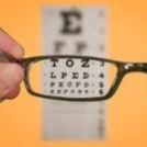
about-author If the cornea is flatter than normal or the eye is short, rays of light are focused behind the retina and causes hyperopia or farsightedness where close objects appear blurred. Astigmatism is a condition in which the uneven curvature of the cornea blurs and distorts both distant and near objects. The cornea is more curved in one direction than in the other. This causes the rays of light to focus on two separate areas of the retina, distorting the visual image. Refractive errors are usually corrected by eye glasses or contact lenses. Although these are safe and effective methods for treating refractive errors, refractive surgeries are becoming an increasingly popular option.

Allergies

about-author

Allergies affecting the eye are relatively common. They are most commonly related to pollen and dust in the air. They are usually immediate or delayed hypersensitivity reactions. Symptoms can include redness, itching, and burning, tearing, stinging and watery discharge. An increasing number of eye allergy cases are related to medications and contact lens wear.


Conjunctivitis (red / pink eye)

about-author

Conjunctiva is a translucent mucous membrane which lines the posterior surface of the eyelids and anterior aspect of the eyeball. A group of diseases that cause swelling, itching, burning and redness of the conjunctiva are termed as conjunctivitis. It is an inflammation of the conjunctiva associated with a discharge which may be watery, mucoid, mucopurulent or purulent. It can spread from one person to another if proper precautions are not taken.

Infections

A breach in the normal epithelial surface of the cornea associated with necrosis of surrounding corneal tissue is termed as corneal ulceration. Two main factors are responsible in the production of a corneal ulcer: damage to corneal epithelium and infection of the eroded area. They usually cause pain, redness, watering, discharge, photophobia i.e. intolerance to light and blurred vision. It can be caused by bacteria, fungi, viruses, acanthamoeba and many other organisms.

Dry Eye

Dry eye can be caused due to any of the following reasons:

  • Aqueous tear deficiency
  • Mucin deficiency
  • Lipid deficiency
  • Impaired eyelid function
  • Corneal epithelium abnormalities.

Corneal dystrophies affect vision in widely differing ways. Some cause severe visual impairment, while a few cause no vision problems and are discovered during a routine eye examination. Other dystrophies may cause repeated episodes of pain without leading to permanent loss of vision.

Keratoconus is characterized by progressive thinning and ectasia which results in deterioration of the quality of vision and also the quality of life. A new modality of treatment, based on collagen crosslinking with the help of Ultraviolet A (UVA, 365nm) and the photosensitizer riboflavin phosphate has been described which changes the intrinsic biomechanical properties of the cornea, increasing its strength by almost 300%.

Pterygia are more common in sunny climates and in the 20-40 age group. Scientists do not know what causes pterygia to develop. However, since people who have pterygia usually have spent a significant time outdoors, many doctors believe ultraviolet (UV) light from the sun may be a factor. In areas where sunlight is strong, wearing protective eyeglasses, sunglasses, and/or hats with brims are suggested. While some studies report a higher prevalence of pterygia in men than in women, this may reflect different rates of exposure to UV light.

Patients with ocular surface diseases suffer from loss of vision, discomfort, infection, erosions, ulceration, and destruction with scarring of the eye surface. The most common cause of these problems is the imbalance in the neural regulation which leads to an “unstable tear film”. Ocular surface failure manifests in two ways – the first one is the Limbal Stem Cell Deficiency in which the corneal epithelium is replaced by the conjunctival epithelium. In the second one, the corneal or the conjunctival epithelium changes with keratinisation and loss of mucosal epithelial characteristics.

A contact lens wearer who presents with conjunctival irritation and associated symptoms – such as itching, burning or tearing – may be suffering from any one of a number of conditions (such as allergy) of which the lenses may not be the primary cause. In other cases, however, contact lens wear itself is responsible for the condition.

Contact lens related problems

A contact lens wearer who presents with conjunctival irritation and associated symptoms – such as itching, burning or tearing – may be suffering from any one of a number of conditions (such as allergy) of which the lenses may not be the primary cause. In other cases, however, contact lens wear itself is responsible for the condition.

Oculoplasty

Ophthalmic plastic surgery or ‘Oculoplasty’ is the branch of ophthalmology that deals not only with the diseases of the eye, but also important structures around the eyes like eyelids, eyebrows, orbit and the tear system which are vital to the normal appearance and function of our eyes – viz Cosmetic Eye Surgery.

Common conditions needing ophthalmic plastic surgery:

  • Eyelid Lift (Blepharoplasty)
  • Entropion
  • Ectropion
  • Eyelid Drooping (Ptosis)
  • Eyelid and Skin Cancer Reconstruction
  • Facial Paralysis (Gold weight implant)
  • Surgery for a Watering Eye DCR
  • Thyroid Eye Disease
  • Orbital Surgery
  • Trauma and Maxillofacial Surgery
  • Pterygium
  • Facial Spasms (Eyelid Spasm) and Botulinum toxin injection


Minimally invasive procedures such as Botox Cosmetic and Filler injections, offer a perfect alternative to cosmetic surgery for busy individuals who can’t afford even a few days of break in their schedules. BOTOX and fillers can achieve temporary yet subtle improvement in signs of aging, without interrupting your day-to-day routine and without incisions, significant swelling or bruising.

  • Blepharoplasty
  • Endoscopic Brow Lift or Forehead Lift
  • Evisceration / Enucleation with customized Prosthesis

Squint Service

Strabismus or Cross-eyes or Squint is a vision problem in which both eyes are not in alignment with each other. Though it is common in infants and children, it can occur at any age. Adults may also develop the condition.

Types of Squint:

  • Intermittent Squint
  • Constant Squint

What Causes Squint

The exact cause of squint is not really known. The movement of each eye is controlled by six muscles. Each of these muscle acts along with its counterpart in the other eye to keep both the eyes aligned properly. A loss of coordination between the muscles of the two eyes leads to misalignment. This misalignment may be the same in all directions of gaze, or in some conditions the misalignment may be more in one direction of gaze, e.g., in squint due to nerve palsy.

Sometimes a refractive error hypermetropia (long sight) may lead to inward deviation of the eye. Poor vision in an eye because of some other eye disease like cataract, etc. may also cause the eye to deviate. Therefore it is important in all the cases of squint, especially in children, to have a thorough eye checkup to rule out any other cause of loss of vision.

  • Decreased in Contrast & Brightness.
  • Due to weak eye muscles or abnormal nerve impulses to the eye muscles
  • Strabismus can accompany some systemic illnesses like diabetes, high blood pressure, multiple sclerosis, myasthenia gravis or thyroid disorders
  • Blurred or poor vision due to cataract, corneal scars, glaucoma, refractive errors, optic nerve disease, retinal disease, tumors of the eye etc.
  • Brought about by injuries
  • Can accompany some systemic illnesses like diabetes, high blood pressure, multiple sclerosis, myasthenia gravis or thyroid disorders.
  • Eyes that look misaligned
  • Eyes that do not appear to move together
  • Frequent blinking or squinting, especially in bright sunlight
  • Tilting head to look at things
  • Faulty depth perception
  • Double vision
  • Poor vision in one or both eyes

The primary goal of treatment is to preserve or restore as much visual function as possible.
Treatment of squint varies depending on the exact type and cause of the strabismus. After a complete eye examination, including a detailed study of the inner parts of the eye, an ophthalmologist can recommend appropriate optical, medical or surgical therapy.
Nearly 40% patients with squint can be cured by spectacles and/or eye exercises. However, a large majority require surgical treatment. The squint operations are very safe and should be done at the earliest. Generally if the eyes are not aligned for more than 6 months in a child, irreversible damage to the three dimensional vision occurs, which is only partly reversible. Squint surgeries are performed successfully even in children as young as 4 months.

Some squints are caused by refractive errors (spectacle powers), usually where one eye has a much higher power than the other. In such cases, squint can be corrected by prescribing proper spectacles.
In children, eye patch may be prescribed for the stronger eye to force the use of the weaker or suppressed eye. Eye drops are used to temporarily blur the vision of the preferred eye. Exercises may be prescribed to strengthen specific eye muscles.
In adult patients double vision caused by small squints can be corrected by incorporating prisms in the spectacles. Squint eye treatment cost varies depending on condition.